Wednesday 27 February 2013

Take the World's Best Courses, Online, For Free


About Coursera

We are a social entrepreneurship company that partners with the top universities in the world to offer courses online for anyone to take, for free. We envision a future where the top universities are educating not only thousands of students, but millions. Our technology enables the best professors to teach tens or hundreds of thousands of students.

Through this, we hope to give everyone access to the world-class education that has so far been available only to a select few. We want to empower people with education that will improve their lives, the lives of their families, and the communities they live in.

Our Courses

Classes offered on Coursera are designed to help you master the material. When you take one of our classes, you will watch lectures taught by world-class professors, learn at your own pace, test your knowledge, and reinforce concepts through interactive exercises. When you join one of our classes, you'll also join a global community of thousands of students learning alongside you. We know that your life is busy, and that you have many commitments on your time. Thus, our courses are designed based on sound pedagogical foundations, to help you master new concepts quickly and effectively. Key ideas include mastery learning, to make sure that you have multiple attempts to demonstrate your new knowledge; using interactivity, to ensure student engagement and to assist long-term retention; and providing frequent feedback, so that you can monitor your own progress, and know when you've really mastered the material.

We offer courses in a wide range of topics, spanning the Humanities, Medicine, Biology, Social Sciences, Mathematics, Business, Computer Science, and many others. Whether you're looking to improve your resume, advance your career, or just learn more and expand your knowledge, we hope there will be multiple courses that you find interesting.

Saturday 23 February 2013

Sudden infant death syndrome


Sudden infant death syndrome (SIDS) is the unexpected, sudden death of a child under age 1 in which an autopsy does not show an explainable cause of death.

The cause of SIDS is unknown. Many doctors and researchers now believe that SIDS is caused by several different factors, including:

Problems with the baby's ability to wake up (sleep arousal)

Inability for the baby's body to detect a buildup of carbon dioxide in the blood


Prevention

Revised American Academy of Pediatrics' (AAP) guidelines, released in October 2005, recommend the following:

Always put a baby to sleep on its back.

Only put babies to sleep in a crib.

Let babies sleep in the same room (NOT the same bed) as parents.

Avoid soft bedding materials.

Make sure the room temperature is not too hot.

Offer the baby a pacifier when going to sleep.

Do not use breathing monitors or products marketed as ways to reduce SIDS.


You can also find some useful guidelines here.

What is also interesting is the Safe to Sleep Public Education Campaign.

The Safe to Sleep campaign—formerly known as the Back to Sleep campaign—aims to educate parents, caregivers, and health care providers about ways to reduce the risk for Sudden Infant Death Syndrome (SIDS) and other sleep-related causes of infant death.


Factors that favor a herniated nucleus pulposus in the L spine


1. Preexisting disc degeneration with tears in the posterior annulus that allow a path for the flow of nuclear material
2. Sufficiently hydrated nucleus capable of exerting high intradiscal pressure
3. Inability of the posterior annulus to resist pressure from the migrating nucleus
4. Sustained or repetitive loading applied over a flexed and rotated spine

source: Donald A. Neumann, Kinesiology of The Musculoskeletal System, Foundations for Rehabilitation

Friday 22 February 2013

Anatomy & Physiotherapy Facebook group


Surprisingly enough, Anatomy & Physiotherapy is a Facebook group that loves sharing useful and evidence based information with its members.

You should definitely be a member: Anatomy & Physiotherapy Facebook group.

About

The latest evidence based conclusions about Anatomy & Physiotherapy on your PC or smartphone App? Follow our Twitter, YouTube, Linkedin and Google+

Description

The latest evidence based conclusions about Anatomy & Physiotherapy on your PC or smartphone App? Follow our Twitter, YouTube, Linkedin and Google+

The importance of pelvic nutation


The relatively small and poorly defined rotational and transnational movements that occur at the sacroiliac joint in the sagittal plane are called nutation and counter-nutation.

These movements perform two major functions, based on Donald A. Neumann, Kinesiology of The Musculoskeletal System, Foundations for Rehabilitation.

1. Stress relief within the pelvic ring
2. Stability during load transfer

Regarding the first function, I have created this flowchart in order to make it understandable:

walking --> reciprocal flexion-extension pattern --> out of phase pelvic rotation --> tension in muscles and ligaments --> oppositely directed torsion --> nutation/counternutation --> stress dissipation (stress that would otherwise occur in the pelvic ring it it were a solid structure).

Regarding the second function, it is important for us to realize that nutation is the closed pack position for the SI joint. This means that the majority of joint structures are under maximal tension in this position, thus the joint is more stable. Consequently, torques that favor nutation help stabilize the SI joint. These are: gravity, stretched ligaments and muscle activation.

Gravity: the body weight line passes vetrally to the sacrum causing a forward tilt & the femural head reaction force on the acetabulutm causes an iliac bacward tilt.

Stretched ligaments: interosseous and sacrotuberous ligaments stretch the joint when movement occurs.

Muscle activation: the following muscle make the sacrum to titl forward (S) or the iliac crest to move backward (I) or just stabilize the SI joint:

erector spinae (S)
multifidus (S)
rectus abd. (I)
obliq. abd. int. & ext.
transv. abd.
hamstrings (I)
gluteus max. (I)
lat. dorsi
iliacus (S)
piriformis

Schober & Ott test for thoracic and lumbar spine RoM


In the method of Schober & Ott, the patient stands erect while the examiner marks the S1 spinous process and a second point 10 cm higher. Regarding the thoracic spine, the examiner marks the C7 spinous process and a second point 30 cm lower (1st figure).

Then the examiner asks the patient to bend as forward as possible and measures the distance between the points again (2nd figure). A normal range of motion (RoM) at the lumbar spine will result in an increased distance of about 15 cm (+5 cm). A normal RoM at the thoracic spine will result in an increased distance of about 38 cm (+8).

Following the same logic, extension in both thoracic and lumbar spine can be assessed (3rd figure).

An alternative method is to measure the smallest finger-to-floor distance with the knees extended.

Revised education policy statement


European Region of the World Confederation for Physical Therapy

Education Policy Statement 

Introduction 

The Executive Committee is pleased to present a revised version of the Education Policy  Statement, prepared by the Education Working Group of the European Region of the WCPT,  taking into consideration the Leuven Communiqué, April 2009.

The purpose of this statement is to provide information to physiotherapists, educators and the appropriate authorities and institutions within the European Union on the principles that underpin programmes of physiotherapy education. These principles cover undergraduate education and the continuum of life-long learning opportunities including higher degrees. 

Central to the statement are the revised WCPT declaration of principle and position statements on Education, the Bologna principles and the direction of the Bologna reforms.  

The Education Policy Statement seeks to ensure excellence in all aspects and levels of education by advocating quality assurance mechanisms. In addition it seeks to inform the work of the European Region in seeking to achieve a European Common Platform by highlighting the need for transparent pathways and  equity in access to physiotherapy education programmes at all levels. The statement aims to provide confidence to the public and to public bodies and the wider higher education sectors promoting the International standing of Physiotherapy Education in Europe.

The entire policy statement is here.

Upper cross syndrome - anatomy


After presenting you my file for the lower cross syndrome, I am now presenting you a similar file for the upper cross syndrome.

A syndrome very common in students that study a lot :-))

Thursday 21 February 2013

International network of spinal cord injury physiotherapists


scipt logo

Who are we?

The International Network of SCI physiotherapists (SCIPT) is a not-for-profit initiative of physiotherapists worldwide. It is for physiotherapists working in the area of spinal cord injuries although other physiotherapists are welcome to join.

What is our mission?

The mission of the International Network of SCI physiotherapists (SCIPT) is to reduce disability and promote participation for people with spinal cord injury and SCI-related disorders (SCI/D) across the globe regardless of income, gender, age, religion, culture or access to health care.


What is within this website?

PowerPoint presentations submitted by members. View the PowerPoint presentations of members. These are presentations members have prepared for conferences, student teaching, program development and the like. Members are free to upload their presentations for others to view. All presentations are categorized for searching purposes.

Sharing of templates, documents and policies. View the assessment and discharge forms other members use in their clinical settings. You will be able to view things such as templates for discharge summaries, goal planning meetings, team meetings, gait assessments etc. In addition, you will find policies written by different organisations on anything to do with SCIPT management.


source: The International Network of SCI

Physiotherapy exercises for people with spinal cord injuries


An amazing tool for physiotherapy exercises for people with spinal cord injuries. You can many different kind of exercises designed for these people and make your own exercise set by filtering them by exercise type, level of injury, difficulty level and other criteria.

Wheelchair rugby


Wheelchair Rugby is a mixed team sport for male and female quadriplegic athletes. A unique sport created by athletes with a disability, it combines elements of rugby, basketball and handball. Players compete in teams of four to carry the ball across the opposing team's goal line. Contact between wheelchairs is permitted, and is in fact an integral part of the sport as players use their chairs to block and hold opponents.

Wheelchair Rugby players compete in manual wheelchairs specifically designed for the sport. Players must meet minimum disability criteria and be classifiable under the sport classification rules. Wheelchair Rugby is a Paralympic sport, with twenty-six countries competing in international competition and more than ten others developing national programs.

Do you find wheelchair rugby soft?


All the hard hits and spectacular crashes at the 2011 Vancouver Invitational Wheelchair Rugby tournament.

Where is your personal limit?


"Bladerunner" Oscar Pistorius raced against a horse in Doha on Wednesday and won - all in an effort to promote disability sport and fight discrimination in the region.

Normally when a horse takes on a human in a race there's usually only one winner but that was before Oscar Pistorius challenged a noble steed to a sprint and it was the horse who came away with a long face.

The Aspire Zone in Doha witnessed "Bladerunner", beating an Arabian horse as part of the 'Definitely Able' campaign which uses special events to highlight the exceptional feats Paralympic athletes are capable of.


Your free resource for physical education and sports coaching


TeachPE.com

Do you wanna be a coach? Or maybe an athlete? Or just study physical education? Here is a wonderful website with a huge amount of information on many kinds of sports, their rules, coaching techniques and a lot of anatomy and physiology required for this science.

TeachPE draws on the expertise and experience of many talented people. To find out more about our experts click on the links below:

Do you know how to sprint..?


Jenny Pacey illustrates how to perform the Skip B drill to help improve sprint technique.

Spine vertebrae range of motion


A very nice visual presentation of the range of motion of each separate inter-vertebrae joint.

Here is the link.

Styled after White AA, Panjabi MM: Kinematics of the spina. In White AA, Panjabi MM, eds: Clinical biomechanics of the spine, Philadelpia, 1990, Lippincott.

Studying tips for the shoulder girdle muscles


It is a bit difficult to learn the muscles, with origin, insertion and action for the shoulder girdle region. One of the most useful methods I have used (suggested by my teacher in anatomy Mr. Bert Loozen) is to separate them in groups and create some kind of mental models, where each model muscles share some common actions and characteristics.

So, regarding the shoulder girdle, the model I follow is the following:

Group 1 - Trunk --> head muscles

1. Sternocleidomastoideus
2. Subclavius

Group 2 - Trunk --> scapula muscles

1. Trapezius
2. Levetor scapulae
3. Serratus anterior
4. Rhombideus major & minor
5. Pectoralis minor

Group 3 - Trunk --> humerus muscles

1. Latissimus dorsi
2. Pectoralis major

Group 4 - Shoulder girdle --> humerus

1. Deltoideus
2. Subscapularis*
3. Supraspinatus*
4. Infraspinatus*
5. Teres minor*
6. Teres major

* rotators cuff muscles

Here is the link with the file i used to study the muscles separated in these group.

Wednesday 20 February 2013

Pelvic floor anatomy & exercises


Maybe we think that pelvic floor muscles are not important in Physical Therapy, but watch these two videos and you will find out how crucial can be for pelvic stability and general health condition.

Gait analysis


One of the most important and interesting things in Physical Therapy is the gait analysis and the analysis of the joints and muscles that take part in it.

I have created a small file that gives a brief overview on the phases of gait and the specific movements of each joint during them.

I think it is helpful for every student.

Three critical elements sustain motivation


Motivating a patient is one of the major parts of the job of a Physical Therapists. So, it is important for him/her to identify the forces that motivate human behavior.

This is a very nice article I recently read in Scientific American:

When it comes to cultivating genius, talent matters, but motivation may matter more.

Got motivation? Without it, the long, difficult hours of practice that elevate some people above the rest are excruciating. But where does such stamina come from, and can we have some, too? Psychologists have identified three critical elements that support motivation, all of which you can tweak to your benefit.

Autonomy

Whether you pursue an activity for its own sake or because external forces compel you, psychologists Edward L. Deci and Richard M. Ryan of the University of Rochester argue that you gain motivation when you feel in charge. In evaluations of students, athletes and employees, the researchers have found that the perception of autonomy predicts the energy with which individuals pursue a goal.

In 2006 Deci and Ryan, with psychologist Arlen C. Moller, designed several experiments to evaluate the effects of feeling controlled versus self-directed. They found that subjects given the opportunity to select a course of action based on their own opinions (for example, giving a speech for or against teaching psychology in high school) persisted longer in a subsequent puzzle-solving activity than participants who were either given no choice or pressured to select one side over another. Deci and Ryan posit that acting under duress is taxing, whereas pursuing a task you endorse is energizing.

Value

Motivation also blossoms when you stay true to your beliefs and values. Assigning value to an activity can restore one's sense of autonomy, a finding of great interest to educators. In a 2010 review article, University of Maryland psychologists Allan Wigfield and Jenna Cambria noted that several studies have found a positive correlation between valuing a subject in school and a student's willingness to investigate a question independently.

The good news is that value can be modified. In 2009 University of Virginia psychologist Christopher S. Hulleman described a semester-long intervention in which one group of high school students wrote about how science related to their lives and another group simply summarized what they had learned in science class. The most striking results came from students with low expectations of their performance. Those who described the importance of science in their lives improved their grades more and reported greater interest than similar students in the summary-writing group. In short, reflecting on why an activity is meaningful could make you more invested in it.

Competence

As you devote more time to an activity, you notice your skills improve, and you gain a sense of competence. In 2006 psychologists at the Democritus University of Thrace and the University of Thessaly in Greece surveyed 882 students on their attitudes and engagement with athletics during a two-year period. They found a strong link between a student's sense of prowess and his or her desire to pursue sports. The connection worked in both directions—practice made students more likely to consider themselves competent, and a sense of competence strongly predicted that they would engage in athletic activity. Similar studies in music and academics bolster these findings.

Carol S. Dweck, a psychologist at Stanford University, has shown that competence comes from recognizing the basis of accomplishment. In numerous studies, she has found that those who credit innate talents rather than hard work give up more easily when facing a novel challenge because they assume it exceeds their ability. Believing that effort fosters excellence can inspire you to keep learning.

The next time you struggle to lace up your sneakers or park yourself at the piano bench, ask yourself what is missing. Often the answer lies in one of these three areas—feeling forced, finding an activity pointless or doubting your capabilities. Tackling such sources of resistance can strengthen your resolve. The choice, of course, is yours.

The Biomotion Lab


BMLwalker


A beautiful tool by "The Biomotion Lab" for "playing around" with different kinds of gait depending on different criteria such as sex, body weight and mood.

The Biomotion Lab

Directed by Prof. Dr. Nikolaus Troje, the lab is located at Queen's University in Kingston, Ontario.

"Goals

We are working on several aspects of visual perception and cognition. Our major interest is focused on questions concerning the biology and psychology of social recognition. That is:

. detection of animate agents
. conspecific recognition
. gender recognition
. individual recognition
. recognition of an agent's actions
. recognition of emotions, personality traits and intentionality
. face recognition".

Monday 18 February 2013

Treatment guidelines: low back pain (2)


I have already presented the treatment guidelines for low back pain from the Royal Dutch Society for Physical Therapy (KNGF).


ACC provides comprehensive, no-fault personal injury cover for all New Zealand residents and visitors to New Zealand.

Sunday 17 February 2013

Village in the Netherlands provides a safe environment for people suffering from dementia


Vivium Zorggroep logo

A village in the Netherlands inhabited entirely by elderly people with dementia offers a new answer to how society can deal with its aging population. It's a world without yesterday or tomorrow where residents have far more freedom than they would be allowed in convalescent homes.


The website of this village is this.

Trunk muscles organized by action


Some of the most difficult muscles to learn in anatomy are the trunk muscles. I just created this file where I put all the trunk muscles organized by action and I thought it would be useful to share it with you.

How to do proper sit ups


There are thousands of books on how to work with your abdominal muscles and which are the best exercises for them. However, only properly educated physiotherapists or trainers should give exercises for the abdominal exercises to people, especially when they are recovering from an injury or a disease.

This article is not about showing more types of exercises, but about explaining how a proper sit up exercise should be performed.

With the body in supine position, 3 major points are about to be analyzed: 1. flexion of the thoracic level, 2. flexion of the knees, 3. reciprocal inhibition of the iliopsoas muscle.

A little bit of anatomy

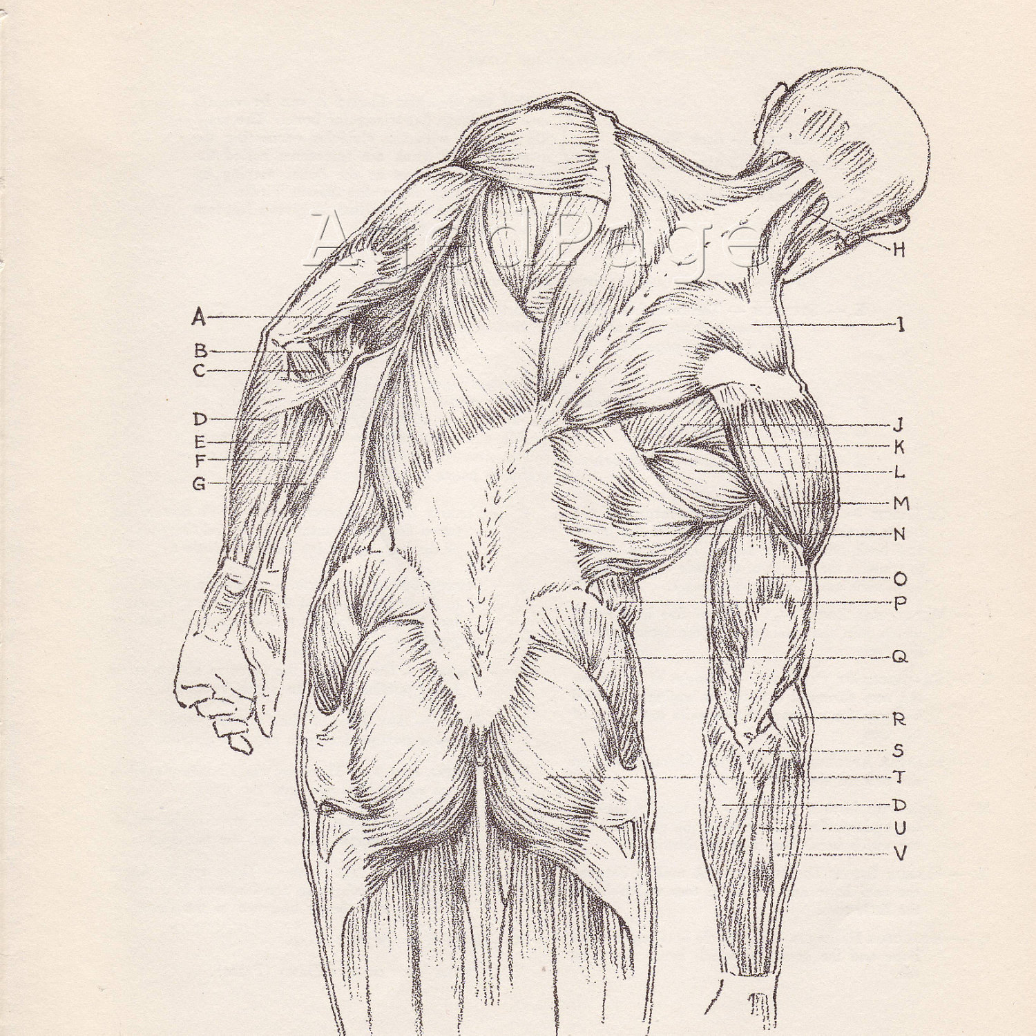
It is important for us to realize the origin and insertion of all our abdominal muscles - the obliquus externus, the obliquus internus, the transversus abdominis, the rectus abdominis,  and the quadratus lumborum (I am not mentioning the pyramidalis, since it does not play a major role in the flexion of the rest of the abdominal muscles).


In the picture above, we cannot see the quadratus lumborum, but this muscle is very deep in the abdominal wall, originating from crista iliaca and inserting to rib 12, and process costalis of L1-L4 vertebrae. You can find more on that in your anatomy book.

The main function of the abdominal muscles is a. the flexion (forward bending) of the trunk against the gravity in the standing posture (our legs fixed) or b. the flexion of our hips in the hanging posture (our trunk fixed). It is very important to realize this "against the gravity" note, because a lot of people thing that when we bend forward while in standing position we use our abdominal muscles, which is not true. When we bend forward while in standing position, it is the gravity that is creating this movement and the back muscles that are activated eccentrically that hold us from falling down.

So, we can understand, that the only way to work with our abdominal muscles is by laying down and start lifting up our trunk with our abdominal muscles against the gravity.

There are also other muscles with the same function - the iliopsoas and the rectus femoris.


It is very important to understand that these muscles are also flexing our trunk (with legs fixed) or flexing our hips (with trunk fixed), because we need to inactivate them and isolate the abdominal muscles when we do the sit ups. In other words, we must be careful when we do sit ups not to work with our iliopsoas or our rectus femoris instead of the abdominal muscles due to wrong posture.

But how we can properly do that? Is it right to lay down completely straight and start doing sit ups? Should we bend our knees? Why? Would it be better to lift them up, above our pelvis? Why? Should we better ask someone to hold our feet on the ground? Or should we better ask him to pull our heels against us while we do the sit up?

Let's try to answer these questions.

In doing the sit ups, as we saw before, we need the isolate the abdominal muscles from the iliopsoas and the rectus femoris. And there are 3 completely safe ways to do that, that every physiotherapist and trainer should be aware of:

1. Flexion of the thoracic level

While doing the sit ups, we should flex only the trunk till the thoracic level, without lifting up the lumbar spine. This way, the abdominal muscles are activated completely. If we try to lift up the lumbar spine, then the iliopsoas is activated and is putting a lot of pressure on the lumbar spine vertebrae, which is quite dangerous, especially in patients with untrained abdominal muscles.

2. Flexion of the knees

By flexing the knees, we bring the biggest percentage of your center of mass above your pelvis or better, above the axis of rotation for the flexion movements of the abdominal muscles. This way, our muscles in the rest of the body are relaxed since we are not trying to hold our legs down on the ground, which we would try to do if we had our legs straight and the center of mass under the axis of rotation for the sit ups. Also, in this posture we rest the lumbar spine my flattening the normal lordosis and putting less pressure on the inter-vertebral disc.

3. Reciprocal inhibition of the iliopsoas muscle

What we must try to do during the sit ups is to inactivate the iliopsoas and the rectus femoris. There are 2 ways to do that:

a. lifting up the legs from the ground. This way, the iliopsoas and the rectus femoris are "loosing" their fixed point (the ground) and they are completely inactivated.

b. bend our knees, keep touching the ground while asking from someone to pull (and NOT hold on the ground) our heels towards him. This way, the gastrocnemius and the hamstrings become active and the iliopsoas and rectus femoris become inactive due to the reciprocal inhibition effect.

So, next time someone asks you to hold his/her feet on the ground while he/she is doing sit ups, do NOT do it :-)

Saturday 16 February 2013

Lower cross syndrome - anatomy


Ok, we all know this diagram. What I was interested in doing, though, was to find the exact muscles that are important in this syndrome. So, I am presenting them with origin, insertion, action and innervation:

Go to this link.

Friday 15 February 2013

Acland's Video Anatomy


Explore anatomical structures and functions with AclandAnatomy.com. This Video Atlas presents expertly dissected human specimens as three-dimensional objects—just as they appear in the living body. Intelligent search and navigation tools make it easy to find the content you need to teach, learn, or review. Ideal for students and instructors as well as practitioners, Acland’s Video Atlas is a virtual anatomy lab at your fingertips.


Propably the most complete Video Atlas..!

Assessment for thoracic outlet syndrome


A very nice video with the most common assessment test for thoracic outlet syndrome:

Observation, Palpation of Scalenes or Cervical Rib
Adson's Test
Reverse Adson's Test
Wright's Test
Eden's Test
Roo's Test

Lumbar spine assessment - a complete guideline


One of the most complete online guidelines for the lumbar spine assessment, broad to us by 3 ESP students of the Hogeschool van Amsterdam - Julie and her two Sara’s.

"The goal of the website is to guide students during the assessment of certain lumbar spine conditions. Therefore, we advise you to first take a look at the anatomy of the lower back to understand the conditions and gain sufficient knowledge on the subject.

The examination page talks about the general examination of the low back, including general patient history, observation as well as active and passive range of motion (ROM) and neurological testing.  This page insures you get the general knowledge you will need to assess a patient with low back problems.

Next to a-specific low back pain and malingering, the website includes lumbar spinal stenosis (LSS), disc herniation, lower crossed syndrome, lumbar instability and scoliosis as low back pathologies. All these conditions are described and subdivided into the different examination parts: patient history, assessment and treatment. The first two subcategories tell you what you should look for/what you might find in a patient with that specific pathology; questions you should ask, what tests to do and what the findings might be. Also in the assessment parts, different assessment tests will be mentioned. When clicking on the test, a link will forward you to the video of the test. The video will show how to perform the test with explanations. The treatment part is only a treatment suggestion and should only be taken as an orientation".

Advanced Massage Techniques


A very helpful YouTube channel with a lot of videos with massage techniques and assessment tests. Broad to us by the Advanced Massage Techniques School.

As the name implies, Advanced Massage Techniques School aims to deliver courses that take students to the highest level in their profession.

Founded in 2009, the School runs courses in the Edinburgh area, providing Diploma courses in:

Orthopaedic Massage and Manipulation
Advanced Remedial Massage
Remedial and Sports Massage
Joint Diploma in Remedial and Sports and Advanced
Remedial Massage

Spondylolisthesis


What is Spondylolisthesis?

The word spondylolisthesis refers to the anterior slippage of one vertebral body with relation to the one below it. The word is derived from two parts - 'spondylo' means spine and listhesis means slippage. A common reason for this type of slippage is weakness or fracture of the portion of the neural arch that connects the lamina with the pedicle, facet joints, and transverse process. This area is the isthmus or pars interarticularis (area between the bony protrusions which articulate to form the joint). As the bridging element between spinal vertebral bodies, these protruding joints are key to the integrity of spinal segments. isthmic spondylolisthesis results from a spondylolisthetic defect in the pars interarticularis, which may lead to the subluxation of a vertebral body.


source: morphopedics

Thursday 14 February 2013

The MRC scale for measuring muscle strength


The patient's effort is graded on a scale of 0-5:

Grade 5: Muscle contracts normally against full resistance.
Grade 4: Muscle strength is reduced but muscle contraction can still move joint against resistance.
Grade 3: Muscle strength is further reduced such that the joint can be moved only against gravity with the examiner's resistance completely removed. As an example, the elbow can be moved from full extension to full flexion starting with the arm hanging down at the side.
Grade 2: Muscle can move only if the resistance of gravity is removed. As an example, the elbow can be fully flexed only if the arm is maintained in a horizontal plane.
Grade 1: Only a trace or flicker of movement is seen or felt in the muscle or fasciculations are observed in the muscle.
Grade 0: No movement is observed.

Monday 11 February 2013

How can you transfer patients with less effort?


Transferring a patient either in a health care unit or in his/her home is the major activities of a Physical Therapist. It is of vital importance for Physical Therapists to learn how to properly transfer patients in many different conditions in order to protect the health of both the patient and themselves.

Here is a beautiful guideline created by WorkSafeBC.

Are you working safely?


"WorkSafeBC is dedicated to promoting workplace health and safety for the workers and employers of this province. We consult with and educate employers and workers and monitor compliance with the Occupational Health and Safety Regulation.

In the event of work-related injuries or diseases, WorkSafeBC works with the affected parties to provide return-to-work rehabilitation, compensation, health care benefits, and a range of other services".

source: WorkSafe BC

They also have a beautiful YouTube channel with very useful videos here.

The UK FIM+FAM (Functional Assessment Measure)


The Functional Independence Measure (FIM) is an 18-item global measure of disability. Each item is scored on 7 ordinal levels. The FIM can be used for measuring disability in a wide range of conditions. The Functional Assessment Measure does not stand alone but adds 12 FAM items to the FIM, specifically addressing cognitive and psychosocial function, which are often the major limiting factors for outcome in brain injury. Hence the Functional Assessment Measure is abbreviated to (FIM+FAM) FAM items are rated on the same 7-level scale as the FIM items although the scaling structure of the FIM does not always lend itself to the more abstract nature of the FAM items.

You can find more information about these methods by visiting the link below:

source: The British Society of Rehabilitation Medicine

The Centre for Evidence-Based Medicine


The Centre for Evidence-based Medicine was established in Oxford as the first of several UK centres with the aim of promoting evidence-based health care. The CEBM provides free support and resources to doctors, clinicians, teachers and others interested in learning more about EBM. Our work falls into three categories: 

1. Research & development on the barriers to and improvement of the clinical practice of evidence
2. Training of students and clinicians in the principles and practice of evidence-based medicine
3. Training the trainers in how to teach EBM and how to do research and development in EBM

source: The Oxford's University Centre for Evidence-based Medicine

Prevention of hospital-acquired infections


A nosocomial infection — also called “hospital-acquired infection” can be defined as:

An infection acquired in hospital by a patient who was admitted for a reason other than that infection. An infection occurring in a patient in a hospital or other health care facility in whom the infection was not present or incubating at the time of admission. This includes infections acquired in the hospital but appearing after discharge, and also occupational infections among staff of the facility.

Patient care is provided in facilities which range from highly equipped clinics and technologically advanced university hospitals to front-line units with only basic facilities. Despite progress in public health and hospital care, infections continue to develop in hospitalized patients, and may also affect hospital staff. Many factors promote infection among hospitalized patients: decreased immunity among patients; the increasing variety of medical procedures and invasive techniques creating potential routes of infection; and the transmission of drug-resistant bacteria among crowded hospital populations, where poor infection control practices may facilitate transmission.

source: the World Health Organization

Sunday 10 February 2013

Cracking your fingers... does it harm your joints..?


Cracking or popping of joints is the action of joint manipulation to produce a sharp cracking or popping sound. This commonly occurs during deliberate knuckle-cracking. It is possible to crack many joints, such as those in the back and neck vertebrae, hips, wrists, elbows, shoulders, toes, ankles, knees, jaws, and the Achilles tendon area.

The physical mechanism causing a cracking sound produced by bending, twisting, or compressing joints is uncertain. Suggested causes include:

1. Cavitation within the joint—small cavities of partial vacuum form in the fluid and then rapidly collapse, producing a sharp sound.
2. Rapid stretching of ligaments.
3. Intra-articular (within-joint) adhesions being broken.

source: Wikipedia

Do you want to find out if cracking your fingers can cause osteoarthritis?


So... now you can continue enjoying cracking your joints..!

Thoracic outlet syndrome


Thoracic outlet syndrome

Thoracic outlet syndrome is a rare condition that involves pain in the neck and shoulder, numbness and tingling of the fingers, and a weak grip. The thoracic outlet is the area between the rib cage and collar bone.

Causes, incidence, and risk factors:

Blood vessels and nerves coming from the spine or major blood vessels of the body pass through a narrow space near the shoulder and collarbone on their way to the arms. As they pass by or through the collarbone (clavicle) and upper ribs, they may not have enough space.

Pressure (compression) on these blood vessels or nerves can cause symptoms in the arms or hands. Problems with the nerves cause almost all cases of thoracic outlet syndrome.

Compression can be caused by an extra cervical rib (above the first rib) or an abnormal tight band connecting the spinal vertebra to the rib. Patients often have injured the area in the past or overused the shoulder.

People with long necks and droopy shoulders may be more likely to develop this condition because of extra pressure on the nerves and blood vessels.

source: U.S. National Library of Medicine


You can also find very useful information on general entrapment neuropathy in both the upper and lower extremities here.

Measuring and using crutches


"Walking aids are used by a variety of people. This includes people recovering from injury, people with  reduced balance strength and endurance, or people with pain or instability in any weight bearing joint used in walking.

The choice of walking aid should suit a person’s abilities and personal requirements. It is recommended to seek the assistance of a physiotherapist in this process.

A physiotherapist is also able to provide advice and training in the correct use of the device.

LifeTec Queensland is a leading provider of information, consultation, and education on assistive technology that can help individuals improve their quality of life and remain independent.

In partnership with a range of complementary organisations, LifeTec Queensland provides advice on the range of available solutions regardless of a person's age or level of ability.

LifeTec assists a wide range of people from all walks of life".


Saturday 9 February 2013

How to locate major vertebrae during assessement


Here are some tips for locating major vertebrae while assessing a patient:

C1 - No proc. spinosus
C1/C2  - lower border upper teeth
C4/C5  - prom. laryingeal
C6 - cricoid, moves most ventrally during extension
C7  - vertrebra prominens
T2/T3 - suprasternal plane
T3  - spina scapulae (trigonum scapulae)
T4/T5 - angulus sterni
T7 - angulus inferior scapulae
T9/T10 - trans. corpus/xyphoid proc.
L3 - subcostal plane
L3/L4 - umbilicus
L4/L5 - crista iliaca
L5 - moves ventrally during extension
S2 - SIPS (lumbalisation/sacralisation)

Source: Anatomy_2 lecture notes, Professor of Anatomy & Kinesiology Bert Loozen (PT - MSc), Hogeschool van Amsterdam

Mayo classification for olecranon fractures


Mayo Classification

The Mayo system describes fractures based on stability, displacement, and comminution.
Type I fractures are undisplaced, type II are displaced and stable, and type III are displaced and unstable. Each is divided into subtype A (noncomminuted) or B (comminuted).

Type I:
Undisplaced fractures: In an undisplaced fracture, it matters little whether a single fragment or several fragments are present; thus, non-comminuted (Type-IA) and comminuted (Type-IB) fractures may be considered to be essentially the same lesion.

Type II:
Displaced, stable fractures: In this pattern, the fracture fragments are displaced more than 3 mm, but the collateral ligaments are intact and the forearm is stable in relation to the humerus. The fracture may be either non-comminuted (Type IIA) or comminuted (Type IIB).

Type III:
Displaced, unstable fractures: The Type-III fracture is one in which the fracture fragments are displaced and the forearm is unstable in relation to the humerus. This injury is really a fracture-dislocation. It also may be either non-comminuted (Type IIIA) or comminuted (Type IIIB).


Go to the source link to see more information on treatment.

Carpal tunnel syndrome


Carpal tunnel syndrome
Median nerve dysfunction; Median nerve entrapment

Carpal tunnel syndrome is a condition in which there is pressure on the median nerve -- the nerve in the wrist that supplies feeling and movement to parts of the hand. It can lead to numbness, tingling, weakness, or muscle damage in the hand and fingers. 


Thursday 7 February 2013

Journal of Orthopaedic & Sports Physical Therapy


"The mission of the Journal of Orthopaedic & Sports Physical Therapy is to publish scientifically rigorous, clinically relevant content for physical therapists and others in the healthcare community to advance musculoskeletal and sports-related practice. Now in its 33rd year, the Journal strives to be the preeminent  source of scientific evidence in musculoskeletal and sports-related rehabilitation and health for the global community.

The monthly JOSPT is the official journal of its publishers, the Orthopaedic Section and the Sports Physical Therapy Section of the American Physical Therapy Association (APTA). Through the print and electronic Journal, the publishers seek to offer high-quality research, immediately applicable clinical material, and useful supplemental information in a variety of formats".

Master in Human Movement Sciences: Sport, Exercise & Health


Application and admission (for foreign students)

Entry requirements
To gain admission to the Master’s Programme Human Movement Sciences you must have a Bachelor of Science degree which includes basic knowledge of Anatomy, Physiology and Psychology on the level of "Introductory textbooks" as well as sufficient research skills and knowledge of Mathematics and Physics. You must also have a good command of English as a working language (TOEFL 580 or IELTS 6,5).

Admission procedure
If you have a Bachelor’s degree and meet all the admission criteria, you can send information to the Admission board of the Faculty of Human Movement Sciences, who will decide if you are indeed admissible.

Residents from countries other than the Netherlands (both within and outside the European Economic Area) have to apply for admission by April 1, 2013. Students who want to be admitted into the Master's programme can apply by filling in the appropriate forms and sending them in together with the required documents. See the VU website for the complete admission procedure.

The 4 major vital signs


Every health professional should know at least the 4 major vital signs:

1. Body temperature
2. Pulse rate (or heart rate)
3. Blood pressure
4. Respiratory rate

1. Body temperature

Body temperature is the degree of body temperature and represents the balance between the produced and dissipated heat of the body.

The purpose of recording the body temperature is to assess and monitor the condition of the patient.

Normal rates:

Body Temperature 36,2 - 37,2 C
Armpit temperature 36 - 37 C
Rectal temperature 36,5 - 37,5 C

2. Pulse rate

Pulse is the wave produced in peripheral arteries when blood enters the aorta after the contraction of the left ventricle. The wave disappears in arterioles before reaching the capillaries.

The purpose is to assess and monitor the condition of the patient. Palpation of the pulse gives us information about the overall patient's situation, especially for arrhythmia, tachycardia, bradycardia.

Normal rates:

For adult 70 to 80 / 1 minute

More than 100 beats per minute is considered as tachycardia.
Below 60 beats per minute is considered as bradycardia.

Where to measure the pulse:

Radial artery, temporal artery, carotid artery, brachial artery, femoral artery, popliteal artery, dorsal artery of foot, posterior tibial artery.


3. Blood pressure

Blood pressure is the pressure of circulating blood volume exerted on the artery walls. The normal regulation of blood pressure depends on the volume of blood, the elasticity of blood vessels, blood viscosity, the diameter of the vessels and the elasticity of the heart muscle.

Systolic or maximum pressure is the highest blood pressure in the arterial wall and corresponds to the constriction of the left ventricle and the flow of the blood into the aorta.

Diastolic or minimum pressure is the lowest value of blood pressure and corresponds to the phase of the dilation of the ventricles.

Pathological Factors:

Age, sex, body weight, exercise, stress, anxiety, certain medication, certain pathologies.

Normal rates (systolic, diastolic)

Male 20 to 45 years old
110 - 140 mmHg 70 - 90 mmHg

Women 20 to 45 years old
100 - 130 mmHg 60 - 80 mmHg

Children 4 years old
100 mmHg 65 mmHg

Newborn
80 mmHg 45 mmHg

For elderly people, we can expect a normal range between 120 - 160 mmHg 80 - 100 mmHg due to expected arteriosclerosis.


4. Respiratory rate

Breathing is the uptake and utilization of oxygen and the production and elimination of carbon dioxide from the cells and the body in general.

The frequency of breaths in adults is 14 to 20 breaths / min, while in children it is normally higher.

Breathing is accomplished with breathing movements, the inhalation and exhalation.

Pathological factors:
Age, sex, disease, exercise, stress, environment, certain medications, obesity, talking, crying, laughing, coughing reflex, swallowing etc.

Types of breath

Apnea is the complete suppression of breathing. Immediate treatment with artificial respiration.
Dyspnea is the subjective sensation of difficulty in breathing (shortness of breath).
Tachypnea is common rapid breathing observed in febrile individuals.
Bradypnea is slow breathing. It occurs when there is damage in the respiratory center.

Wednesday 6 February 2013

Do you have a correct posture in your ADLs?


Logo

With a correct posture can prevent physical symptoms. In this website you will find instructions for the correct posture at work in the nursing care and home care.

You have to use google.translate because it is in Dutch, but you can understand everything since it has many many pictures in it.